


Excellent to Assess the Morphological Features of Sperm Cells
Sperm morphology is assessed routinely as part of standard laboratory analysis in the diagnosis of human male infertility. It is well-known that the morphology of fertile sperm is significantly different from that of infertile sperm.
Optical microscopy has been used to observe the morphological features of sperm cells, but requires sample staining and offers only limited resolution.
In contrast, atomic force microscopy doesn’t need any sample staining. Furthermore, it provides much higher resolution images than optical microscopy.
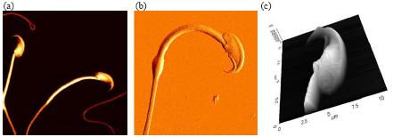
Figure 1. High resolution AFM Images of mouse sperm cells. (a) Topography of sperms (45 μm scan size), (b) error image of sperm (25 μm scan size) and (c) 3D rendering of topography image of sperm head (11 μm scan size).
Figure 1 and 2 show high resolution AFM images of mouse sperm cells. Mouse sperm cells were extracted from the mouse testis tissue and fixed with 1% glutaraldehyde and 1% paraformaldehyde in phosphate buffered saline. Then, they were attached onto the poly-L-lysine coated glass slide and it was washed out with filtered distilled water twice.
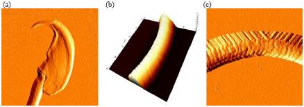
Figure 2. High resolution AFM images of mouse sperm cells. (a) Error image of sperm head (11 μm scan size), (b) 3D rendering of topography image of sperm tail (5 μm scan size) and (c) error image of sperm tail (5 μm scan size).
The images were obtained using contact mode AFM in air. A gold-coated silicon nitride cantilever was used to image the sample. Its spring constant is approximately 0.06 N/m.
AFM images can be quantitatively analyzed using XEI software. The dimensions and features of sperm cell parts can be measured and evaluated with the strict criteria for sperm morphology.
The Park AFM is expected to become an excellent tool to assess the morphological features of sperm cells.