


Special Modes
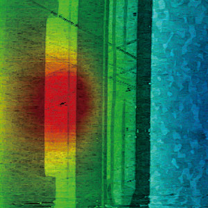
Scanning Thermal Microscopy (SThM)
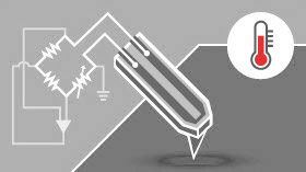
High Spatial and Thermal Resolution Microscopy
Our SThM mode lets you easily find the local thermal conductivity of a sample by measuring heat transfer between tip and sample using a micro-fabricated probe.
Read More
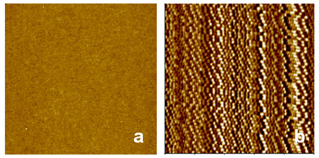
Magnetic Force Microscopy (MFM)
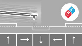
High Resolution and High Sensitivity Imaging of Magnetic Properties
Our MFM mode measures the magnetic variations over a sample surface by detecting the interaction between a magnetized cantilever and sample surface. The cantilever measures surface topography on the first scan, then lifts and follows either the stored surface topography (lift mode, available only in selected countries) or a constant distance (or constant height) at a fixed height above the sample surface.
Read More
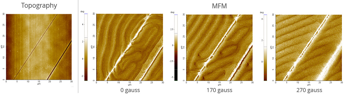
Tunable Magnetic Field MFM (TM-MFM)
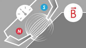
High Resolution and High Sensitivity Imaging of Magnetic Properties
Our TM-MFM mode measures the magnetic domain distribution with respect to magnetic field change.
Chemical Force Microscopy (CFM)
CFM measures the chemical interactions between functionalized tips and sample to determine the chemical nature of surfaces and facilitate studies of chemical bonding enthalpy and surface energy. Tips are typically gold-coated and functionalized with R-SH thiols, R being the functional groups of interest such as -CH, -COOH, -NH, and -OH.
Electrochemical Microscopy (EC-AFM)
In EC-AFM, STM the nanoscale structures of electrochemical reactions on the electrode surface can be observed. Users typically perform voltammetry and corrosion experiments using an electrochemistry cell and a choice of potentiostat/galvanostat depending on the electrochemical application of interest.
Scanning Ion Conductance Microscopy (SICM)
In Scanning Ion Conductance Microscopy developed by Park Systems (Park SICM), a glass nanopipette (a pipette in the nanoscale) filled with an electrolyte, acts as an ion sensor that provides feedback on its location relative to a sample completely immersed in a liquid. The pipette-tip maintains its distance from the sample by keeping the ionic current constant. In comparison, Atomic Force Microscopy (AFM) typically relies on interaction of forces between a probe tip and the sample.
Scanning ElectroChemical Cell Microscopy (SECCM)
Scanning probe microscopy (SPM), and especially pipette-based SPM techniques can offer new insights into the nano-scale chemical properties of the sample. Among the pipette-based SPM techniques, Scanning electrochemical cell microscopy (SECCM) is a recently developed pipette-based SPM technique designed to investigate the local electrochemical properties of surfaces. Using laser-pulled pipettes with nano or microscale tip radius, SECCM allows investigating electrochemical responses on target materials by applying a bias sweep, also known as cyclic voltammetry (CV).