


Brian Choi,Bio-application scientist
For more information, please contact app@parksystems.comSample courtesy: Hong-Bae Kim, Seoul National Univ.
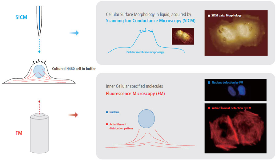
The combination of fluorescence microscopy and SICM techniques can create new benefits and can provide comprehensive information for cell biology studies that is not obtainable when using only one of those techniques. While monitoring the external cellular surface morphology with SICM, the internal cellular behavior can be observed by fluorescence microscopy. In this study, an electroporated H460 cell in buffer was observed using SICM and fluorescence microscopy. The irreversible electroporation was performed at 1 kV electric pulse, and the cell was fixed with 4 % paraformaldehyde. Then the cell was imaged with SICM imaging in 1x PBS. After staining the cell with rhodamine phalloidine (red) for actin filament identification in the cell, fluorescence microscopy images were captured.
- More Comprehensive Cell Biology Study by Integrating Fluorescence Microcopy with SICM
- While monitoring external cellular surface morphology with SICM, the internal cellular molecule can be observed by Fluorescence microscopy
Simultaneous Cell Observation of SICM (surface morphology) and Fluorescence Microscopy (actin filament structure) for Fundamental Cell Analysis
SICM imaged the treated H460 cell membrane topography first, and then the actin filament and nucleus images were acquired using fluorescence microscopy. The SICM image shows that the plasma membrane of H460 cell was physically damaged by electric pulse and many holes appeared on the surface. In the fluorescence microscopy image, the network structure of actin filaments inside the cell was clearly detected but were not damaged much.
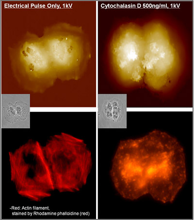
Cytochalasin (500 ng/㎖ ) treatment on H460 cell resulted to the same irreversible electroporation. Cytochalasin causes the actin filament to break off suddenly, by binding on it and blocking the polymerization. This action of cytochalasin on actin filament was distinctively detected by fluorescence microscopy observation. Interestingly, however, cytochalasin treated H460 cell does not show any holes on the cell membrane.
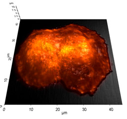 Image Overlay
Fluorescence Microscopy Integration with SICM for Cell Study
Park Cell Analysis Systems
|
|
|
|
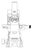
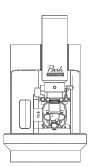
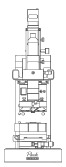
| Park NX12-Bio | Park NX10 | Park XE7 | |
| Scanning Ion Conductance Microscopy (SICM) | |||
| Atomic Force Microscopy (AFM) with liquid probe hand | |||
| Inverted Optical Microscopy (IOM) | |||
| Live Cell Chamber |