
The DPG Spring Meeting of the Condensed Matter Section (SKM) will take place on the University of Regensburg's campus from March 16 to 21, 2025. Park Systems will be one of around 100 companies taking part in the Exhibition of Scientific Instruments and Literature from Tuesday to Thursday.
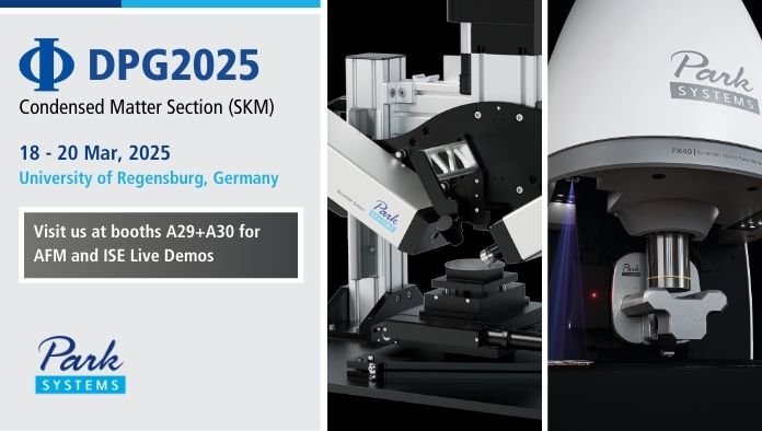
From revolutionary automation features for nanoscale imaging to high-resolution measurements, we will have Park Systems demo tools available at booth A29 + A30! Join us for a live demonstration on:
• Accurion Simon Imaging Ellipsometer, with a lateral resolution of 1 µm.
• The FX40 Automatic AFM, with built-in AI and machine learning.
- Date: 18 – 20 March, 2025
- Venue: University of Regensburg, Germany
- Booth#: A29 + A30
Link: https://regensburg25.dpg-tagungen.de/
Join our talk on Friday, March 21, 2025, 11:00–11:15, H34 in Session CPP 40: Energy Storage and Batteries II.
Title: Measuring Local Electrochemical Properties with Scanning Probe Microscopy
Presenter: Dr. Alexander Klasen, Principal Scientist, Park Systems Europe
Co-Author: Dr. Andrea Cerreta, Principal Scientist, Park Systems Europe
Abstract:
Electrochemical (EC) applications, ranging from novel energy storage systems to advanced catalysts, are defined on an ever-decreasing length scale. Investigating these systems requires to map key functional features with sufficient resolution, such as the local structure, electronic properties and electrochemical response. Scanning probe microscopy-based techniques are well established to investigate surface parameters using the physical interaction with a nanometer-sized probe allows studying properties such as the topography, work function, or adhesion at high resolution.1,2
One such technique, Scanning Electrochemical Cell microscopy (SECCM), was first introduced by E. Daviddi, P.R. Unwin, et al. and uses a pipette-based SPM approach to probe local EC features. 3
When brought in close proximity to the sample, the electrolyte-filled pipette pipette creates a small electrolyte meniscus between the pipette aperture and the surface of interest. This confined volume of solution constitutes a small electrochemical cell that allows for local measurements of electrochemical characteristics.
In this talk, we discuss the basics of SECCM, present recent examples from literature, discuss the limitations of the technique and outline potential pathways to overcome those.4–7
1. Eaton, P. & West, P. Atomic force microscopy. (Oxford university press, 2010).
2. Wiesendanger, R. Scanning probe microscopy and spectroscopy: methods and applications. (Cambridge university press, 1994).
3. Daviddi, E., Gonos, K. L., Colburn, A. W., Bentley, C. L. & Unwin, P. R. Scanning Electrochemical Cell Microscopy (SECCM) Chronopotentiometry: Development and Applications in Electroanalysis and Electrocatalysis. Anal. Chem. 91, 9229–9237 (2019).
4. Liu, G. et al. A simple approach for effectively improving the resolution of scanning electrochemical cell microscopy. Sensors Actuators B Chem. 409, 135603 (2024).
5. Choi, M. et al. Probing Single-Particle Electrocatalytic Activity at Facet-Controlled Gold Nanocrystals. Nano Lett. 20, 1233–1239 (2020).
6. Brunet Cabré, M., Paiva, A. E., Velický, M., Colavita, P. E. & McKelvey, K. Electrochemical kinetics as a function of transition metal dichalcogenide thickness. Electrochim. Acta 393, (2021).
7. Cabré, M. B., Paiva, A. E., Velický, M., Colavita, P. E. & McKelvey, K. Electrochemical Detection of Isolated Nanoscale Defects in 2D Transition Metal Dichalcogenides. J. Phys. Chem. C 126, 11636–11641 (2022).