



Ovidiu-I. Agârbicean is a third-year undergraduate student in Medical Physics at Babeș-Bolyai University, Cluj-Napoca, Romania, with a strong interest in theoretical and medical physics. His work focuses on bridging theoretical models and experimental investigations, applying concepts from statistical physics and interfacial physics to experimentally driven problems. He is particularly interested in connecting fundamental physical principles with measurable phenomena, fostering a closer integration between theoretical understanding and experimental evidence.
1. Please summarize the research you do and explain why it is significant?
My research focuses on developing a theoretical framework for understanding potential-dependent adsorption and desorption at the solid–liquid interface of citrate-stabilized silver nanoparticles (AgNPs), and how these processes influence surface-enhanced Raman spectroscopy (SERS).
SERS can provide highly sensitive molecular detection, but its response depends on more than the optical properties of the nanoparticles. The surface charge, electronic state, local chemical environment, and adsorption of the target molecules can all influence the measured signal.
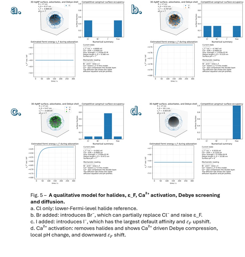
My work investigates how changes in the electrochemical potential of AgNPs can modify their surface chemistry. In particular, I study the effects of Ca²⁺ addition, which can promote surface Ag⁺ formation and alter the interfacial environment, and halide adsorption, which can modify the electronic state of the nanoparticles. These changes can favor or suppress the adsorption of particular molecular species and therefore influence their SERS response.
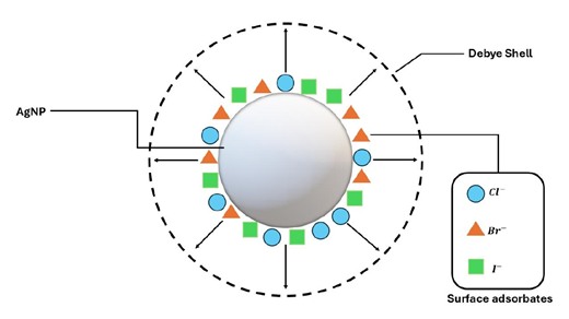
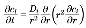
To investigate the transport processes near the nanoparticle surface, I use the spherical radial diffusion equation as a first approximation, neglecting migration and convection under conditions where their contributions can reasonably be considered small. I am also interested in how differences between proton and hydroxide transport could contribute to local pH gradients near the interface.
The significance of this research is that it aims to connect statistical physics, ion transport, surface electrochemistry, and molecular spectroscopy within one framework. Rather than interpreting every change in SERS intensity as a consequence of nanoparticle aggregation, I investigate whether changes in adsorption and desorption can provide an alternative or complementary explanation.

Ultimately, I hope this approach will help establish more predictable relationships between nanoparticle surface conditions, molecular adsorption, and optical signals.
2. How might your research be used?
One potential application is the development of more controllable, label-free optical sensing platforms for chemical and biological detection.
SERS can provide molecularly specific vibrational fingerprints, but the sensitivity and reproducibility of a measurement often depend on how analytes interact with the nanoparticle surface. If we can understand and control how surface potential, ionic composition, and pH influence adsorption, we may be able to improve the conditions under which a target molecule produces a detectable Raman signal.
This is particularly relevant to biomarker detection. Many biologically relevant molecules have different charges and chemical functionalities, so controlling the nanoparticle–molecule interface could help favor the adsorption of a particular target or reduce interference from competing species.
For example, my work on citrate-stabilized AgNPs investigates how Ca²⁺ and halide ions alter the surface environment and electronic state of the nanoparticles. The broader idea is to use these interfacial changes as a means of controlling molecular adsorption and, consequently, the optical response.
At this stage, I see this primarily as a fundamental research direction rather than a validated diagnostic method. The next steps would involve testing the model against experimental measurements and evaluating whether the resulting control over adsorption improves sensitivity, selectivity, and reproducibility in more complex samples.
3. Why is the Park AFM important for your research?
The Park AFM is particularly relevant to my research because it can help connect the physical structure of a nanoparticle system with its spectroscopic response.
My theoretical framework considers how surface potential, adsorption, and the local chemical environment influence SERS. However, when a Raman signal changes, it is important to determine whether the change is associated with molecular adsorption, nanoparticle aggregation, changes in surface morphology, or a combination of these effects.
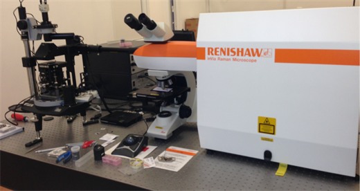
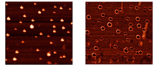
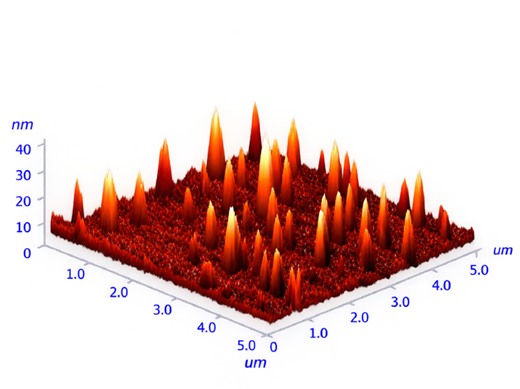
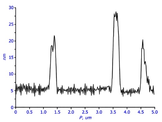
Atomic force microscopy can provide information about surface topography, particle dimensions, and the spatial organization of deposited nanoparticles. When combined with Raman spectroscopy in an integrated Raman-SPM system, it becomes possible to correlate structural information with spectroscopic measurements.
This would be valuable for testing my hypotheses experimentally. For example, I could compare nanoparticle samples before and after Ca²⁺ or halide addition, examine their morphology, and investigate whether changes in the Raman response correlate with observable structural changes.
The combination is especially interesting because it could help distinguish structural effects from changes associated with molecular adsorption and interfacial chemistry. It would therefore provide an experimental route to test and refine the theoretical model, rather than relying on Raman intensity alone.
4. What features of Park AFM are the most beneficial and why?
For my research, I would prioritize three capabilities: high-resolution topographic imaging, integration with Raman spectroscopy, and spatially correlated measurements.
First, high-resolution AFM imaging would help characterize the morphology and distribution of nanoparticles deposited on a substrate. This is important because nanoparticle size, particle–particle spacing, and aggregation can influence the local electromagnetic field and therefore the measured SERS intensity.
Second, the integration of AFM with Raman spectroscopy would allow structural and spectroscopic information to be collected from the same sample and, where supported by the instrument, from corresponding locations. This would help me investigate whether a change in the Raman signal is associated with a structural modification or is more consistent with a change in the molecular environment at the surface.
Third, the ability to examine spatial variations would be useful for studying heterogeneous nanoparticle surfaces and identifying regions with different topographic or spectroscopic characteristics. Such measurements could help evaluate the reproducibility of SERS signals and determine whether the response is dominated by a small number of highly active regions.
I would also value reliable imaging modes, stable measurements, and the ability to work with different sample types, depending on the configuration available. Together, these capabilities would help me connect theoretical predictions about interfacial processes with experimental evidence at the nanoscale.


